0:37, silent. Watch it with music on Instagram.
How small is the deep brain stimulation target?
The subthalamic nucleus is about the size of a coffee bean. Here are the target, the electrode and the brain, all to scale.
- A whole brain is about 16 cm long.
- The target, the subthalamic nucleus (STN), is a lens-shaped nucleus about 1 cm long and 3–5 mm thick: about the size of a coffee bean.
- The DBS lead is about 1.3 mm wide (1.27 mm for the common 4-contact leads), with contacts 1.5 mm tall. It goes in through a small hole in the skull and travels about 8 cm through the brain to the target (in this model).
- Surgeons aim at its motor (dorsolateral) part, where stimulation best eases the motor symptoms.
- Typical placement error is about 2 mm (pooled studies), so millimetres decide the result.
- Clinical: a few millimetres off, the current reaches a neighbour (seen from above):
- too lateral: the internal capsule, muscle pulling (often in the face) and slurred speech
- too medial: the oculomotor nerve fibres and the limbic part of the STN, double vision and mood changes (e.g. hypomania)
- too posterior: the medial lemniscus, tingling
- too anterior: the hypothalamus, sweating and nausea
Too deep, into the substantia nigra, can lower the mood. Programming (or a directional lead) can often steer the current away.
DBS does not cure Parkinson's or stop it, but in trials it improved off-medication motor scores by about 40% and roughly halved the medication. Typical stimulation: ~130 pulses a second.
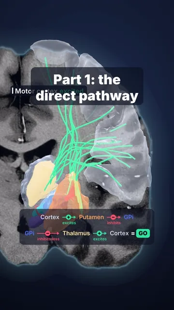
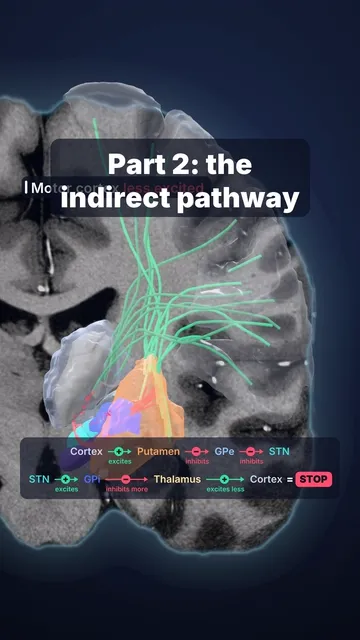
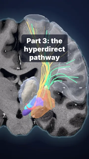
Part 3 of our basal ganglia series showed why the STN matters: it is the brain's emergency brake.
Sources and licence
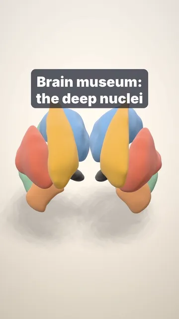
Data: cortex and deep nuclei from the 7 T MRI of one volunteer (Lüsebrink et al., Sci Data 2021, CC0); subthalamic nucleus, substantia nigra, red nucleus and internal capsule from the MASSP 2.0 atlas (Alkemade et al., CC BY 4.0); mammillary body (hypothalamus) from the Neudorfer et al. 2020 hypothalamic atlas (CC BY 4.0); the lead is drawn to scale (schematic trajectory); the medial lemniscus and the oculomotor fibres are named, not drawn. Models in MNI template space. Facts: Pasricha 2023 and Mourgela 2007 (brain length); Schäfer 2012 (STN size); Krauss et al., Nat Rev Neurol 2021 (leads); Dembek et al.
Educational content, not medical advice.