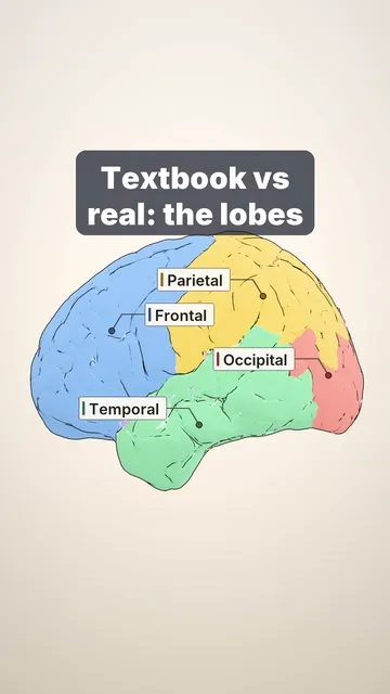
0:34, silent. Watch it with music on Instagram.
Two language areas and the cable between them
Why can a stroke take away your words but not your understanding, or the other way round?
The classic language model (left hemisphere in most people):
- Broca's area (inferior frontal gyrus) plans speech. Damage: slow, effortful, halting speech, while understanding is relatively spared (Broca's aphasia).
- Wernicke's area (back of the superior temporal gyrus) helps understand words. Damage: fluent speech that makes little sense, and poor understanding (Wernicke's aphasia).
- The arcuate fasciculus is the white-matter cable between them. Damage: trouble repeating words (conduction aphasia).
Real language networks are bigger (more areas, more pathways), but this model is still the exam classic.
Sources and licence
Rendered from open data: lobes, gyri and 7 T MRI of a volunteer (Lüsebrink et al., Sci Data 2021, CC0); arcuate fasciculus from the HCP1065 atlas (Yeh, Nat Commun 2022, CC BY-SA 4.0), data provided by the Human Connectome Project, WU-Minn Consortium (Principal Investigators: David Van Essen and Kamil Ugurbil; 1U54MH091657) funded by the 16 NIH Institutes and Centers that support the NIH Blueprint for Neuroscience Research; and by the McDonnell Center for Systems Neuroscience at Washington University. Wernicke's area border and signal dots drawn schematically.
Video licence: CC BY-SA 4.0. To reuse it, credit “Cerebrarium (Aldo Spolaore)”, link to this page and name the data sources above.
Educational content, not medical advice.